Understanding Facebows in Dentistry
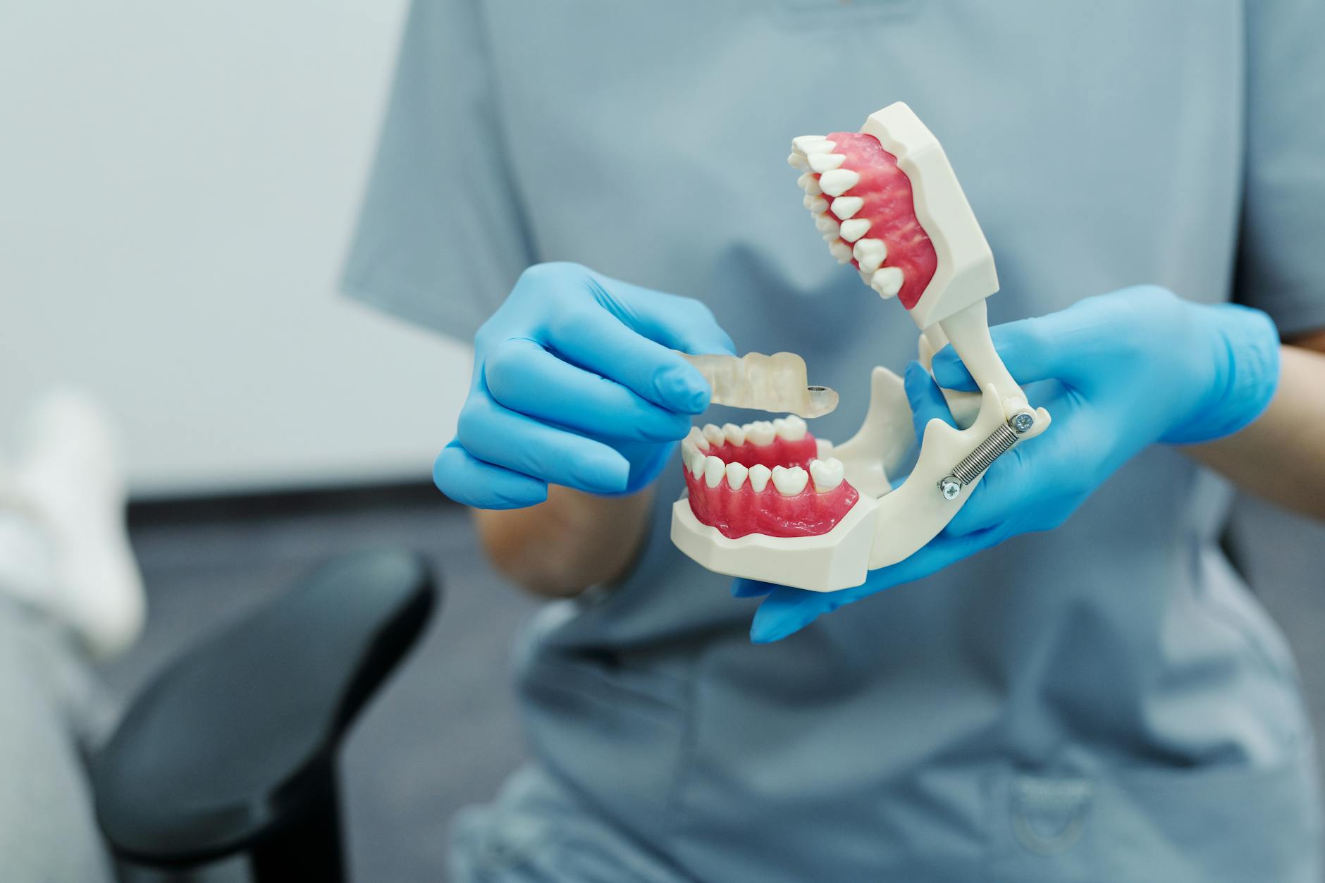
A facebow is a device dentists use to record how your upper jaw sits in relation to your skull and jaw hinge. That record lets us transfer your mouth’s position to a jaw simulator (articulator) so planned teeth match your bite and facial features. It matters most when small alignment errors could affect comfort, function, or appearance.
How it works: an earbow or a kinematic facebow references facial landmarks (often near the ears) and a head plane to orient a maxillary model on an articulator. That orientation helps guide the occlusal plane, midline, and tooth display during chewing and smiling. For multi‑unit cases, this record supports tasks like planning precise crowns and bridges. Studies comparing arbitrary transfer devices with a true kinematic facebow show measurable differences in mounted cast position [1]. Other research has compared dento‑facial analyzer systems with earbow records [2].
Today, digital workflows can capture facial information with photos or a 3D face scan and merge it with intraoral scans in software. This lets the team align the virtual occlusal plane and smile line to your facial features, then design restorations such as thoughtful smile design with porcelain veneers. In conversations about facebow vs digital face scan esthetics, both aim to place teeth where they function well and look natural; the tools differ, but the goal is the same. Reviews describe how newer devices and virtual articulators transfer maxillary orientation in the digital space [3].
A facebow or its digital equivalent is especially useful when:
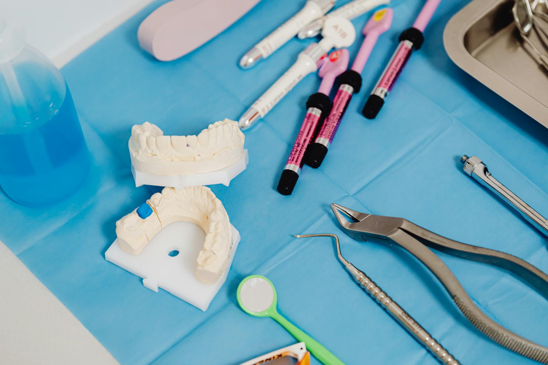
- Planning a full‑mouth rehabilitation or major bite change
- Making complete or partial dentures
- Designing multiple connected crowns or bridges
- Rebuilding worn, tilted, or uneven bite planes
- Coordinating complex implant restorations with jaw movement
Have questions about visits or timing? See our current hours.
The Evolution to Digital Face Scans
Digital face scans are a modern way to capture how your teeth and smile relate to your face. They have largely replaced or complemented traditional facebows by recording a 3D image of your face and merging it with your mouth scan. This helps orient the bite and smile line in software so planned teeth look natural and work well.
What changed is speed, detail, and how the data connects. A calibrated camera or photo set captures your facial landmarks and head posture in seconds. Software then aligns that 3D face with an intraoral scan to set the occlusal plane and midline, and uses a virtual articulator to preview jaw movements. That virtual setup guides design for treatments ranging from single crowns to full smile makeovers and clear aligners, improving communication with the lab and with you. It also streamlines planning for precise, staged tooth movement in clear aligner treatment planning.
Both methods aim for the same goal: put teeth where they function well and look like they belong to your face. A facebow records jaw position with physical landmarks; a digital face scan records the same relationships visually and integrates them directly into design software. In the facebow vs digital face scan esthetics discussion, the choice often comes down to workflow: digital makes it easier to check symmetry, smile arc, and tooth display against your own 3D image, while a well‑made analog record still works reliably. Either way, clinicians verify the plan by cross‑checking midlines, planes, and bite records before finalizing.
For patients, the process is simple: look toward a point while the camera captures a few images; there is little or no contact, and it takes about a minute. We may capture a brief bite record to sync jaw position to the scan, then preview options with you on screen. This approach is especially helpful when designing dentures or implant restorations, where lip support and smile fullness matter, such as planning snap‑in implant dentures. Your photos and scans are part of your dental record and are used only to plan care.
Comparing Accuracy: Facebow vs Digital Scan
Both methods can accurately orient the upper jaw to the face when used correctly. A traditional facebow aims to locate the hinge axis and transfer the maxilla to an articulator; a digital face scan captures facial symmetry and head posture, then aligns the smile and bite in software. In everyday planning, the practical accuracy for esthetics is similar, but each approach has different sources of error in capture and transfer—so careful verification matters in either case. This balance often guides the facebow vs digital face scan esthetics discussion.
Analog accuracy depends on the type of facebow and how consistently it is used. Kinematic facebows can closely approximate the true hinge axis, while earbows are faster but assume an average axis. Small angular differences during transfer can change the occlusal plane or midline on the articulator. Patient posture, reference plane choice, and how securely the bite record is made also influence results; clinicians often double‑check these with trial mounts and articulated wax‑ups before committing to final restorations.
Digital accuracy relies on correct camera calibration, a stable “natural head position,” and precise registration between the 3D face and the intraoral scan. When those steps are sound, software makes it easier to align the occlusal plane to the interpupillary line, preview smile arc, and visualize lip support. Function is confirmed by synchronizing a consistent bite record or jaw motion data with the virtual setup. This visualization can be especially helpful when designing and fitting esthetic frameworks or clasps for well‑balanced partial dentures.
In full‑arch or complex cases, accuracy improves when we combine methods and verify: mount or virtually align the maxilla, check midline and planes on the face, then test with a mock‑up or printed try‑in. If jaw position is critical, a kinematic facebow or validated digital jaw‑motion record adds confidence; for purely esthetic alignment, a high‑quality face scan with proper registration often suffices. Teams commonly iterate: adjust the plane or incisal display in software, preview with a provisional, then finalize—an approach that works well for coordinated All‑on‑4 full‑arch implant planning.
Benefits of Digital Face Scanning
Digital face scanning helps us place teeth where they look natural on your face and work well with your bite. It captures a precise 3D image of your face and links it to your mouth scan, so we can design with your real smile, lips, and head posture in view. The process is quick, comfortable, and non-contact, and it improves planning and communication with you and the dental lab.
Because the 3D face and intraoral scans are merged in software, we can align the occlusal plane to your eyes, center the midline, and preview tooth display at rest and in a smile before any treatment begins. This makes it easier to test shapes and lengths, adjust symmetry, and show you options on screen. It often reduces try-ins and chairside adjustments, since the plan is checked against your own facial features early. The same workflow supports single-tooth fixes or full smiles, including designing precise, conservative dental bonding for small chips or edges.
Digital scans are also repeatable: we can capture your natural head position the same way at different visits, track changes over time, and store records securely. Compared with only analog records, it is easier to spot small tilts, asymmetries, or lip support needs and correct them in the plan. In the facebow vs digital face scan esthetics conversation, the digital approach adds clear visual context while still allowing a bite record or articulator check when needed. It also helps with shade planning and photo‑matched mock‑ups, including setting a target brightness for thoughtful tooth whitening before restorations.
Quality depends on good steps: a calibrated camera, a steady gaze for natural head position, and a consistent bite record to sync the jaws. When we follow these, the result is a smoother design process, fewer remakes, and restorations that fit your face as well as your bite. Not every case needs a full face scan, but when esthetics or complex tooth positions matter, it gives the team and patient a clearer, shared picture of success.
Impacts on Smile Aesthetics
Both a facebow and a digital face scan aim to place your teeth in harmony with your face. They influence the occlusal plane (the “tilt” of your bite), the midline, tooth display at rest and in a smile, and how the lips frame your teeth. When these are set well, the smile looks natural and balanced; when they are off, the smile can look canted or uneven.
A facebow helps orient the upper jaw on an articulator so tooth length and plane match average head landmarks. This supports even incisal edges, a level smile line, and reduces the chance of a visible cant. A digital face scan captures your actual head posture and facial symmetry, then aligns the virtual teeth to your interpupillary line and smile arc in software. That visual context makes it easier to judge tooth length, buccal corridor fullness, and gingival display before any work is done. In the facebow vs digital face scan esthetics discussion, both can succeed; digital mainly adds a clear picture of your own face to guide choices.
Small changes in incisor inclination, tooth color, and other “micro‑aesthetic” details can shift how a smile is perceived by both dentists and laypeople, so precise orientation matters [4]. Digital smile design methods can improve planning and communication, letting the team and patient preview options and agree on esthetic goals earlier in the process [5]. When we print or fabricate a mock‑up from that plan, studies show that supported indices and standardized trays can improve the reproducibility of these trial restorations, which helps translate the planned look to the mouth more consistently [6].
Function and esthetics go together. Proper orientation supports clear speech sounds, comfortable chewing, and even wear. It also helps set lip support and incisal show that suit your face at rest and in motion. In complex cases, clinicians often verify the plan with a provisional or try‑in, then make small adjustments to plane, midline, or edge position before finalizing. This stepwise approach reduces surprises and helps the final smile look like it belongs to you.
Streamlining Cosmetic Planning
Cosmetic planning moves faster when facial data and tooth data come together at the start. A 3D face scan or calibrated photos are merged with your mouth scan and a stable bite record so we can set the plane, midline, and tooth display in software before any drilling or shaping. This shortens back‑and‑forth with the lab, reduces surprises, and helps everyone see the same plan. In the facebow vs digital face scan esthetics discussion, this shared visual makes it easier to agree on what “natural” should look like for you.
The streamlined workflow is simple: capture your natural head position while you look at a point, take the 3D face or photo set, record the bite, and scan the teeth. The software aligns your smile to your interpupillary line and facial midline, then uses a virtual articulator to preview how the teeth will meet. Because the face and teeth live in one file, we can test tooth length, shape, and symmetry on your own image, save versions, and send the same view to the lab. If a detail looks off, we adjust it on screen and re‑export in minutes.
Compared with a purely analog path, fewer physical transfers means fewer places for small errors to sneak in. A traditional facebow can still be valuable, but it often requires model mounting, shipping, and extra wax steps to see changes on the face. Digital planning keeps the head posture consistent from visit to visit, makes midline and plane checks obvious, and lets us print a mock‑up or provisional directly from the approved setup. That speeds review and keeps the team aligned on the same esthetic and functional targets.
For patients, this translates to clearer choices and usually fewer try‑ins. We can show you options side by side, agree on a target look, and test it with a conservative mock‑up before final work. Complex bites may still benefit from added records—such as a kinematic facebow or jaw‑motion data—while simpler cosmetic goals may need only a high‑quality face scan with a consistent bite record. Either way, starting with your face on screen helps us plan a smile that fits your features and your bite, with a smoother, step‑by‑step path from preview to final.
Patient Experience with Digital Scans
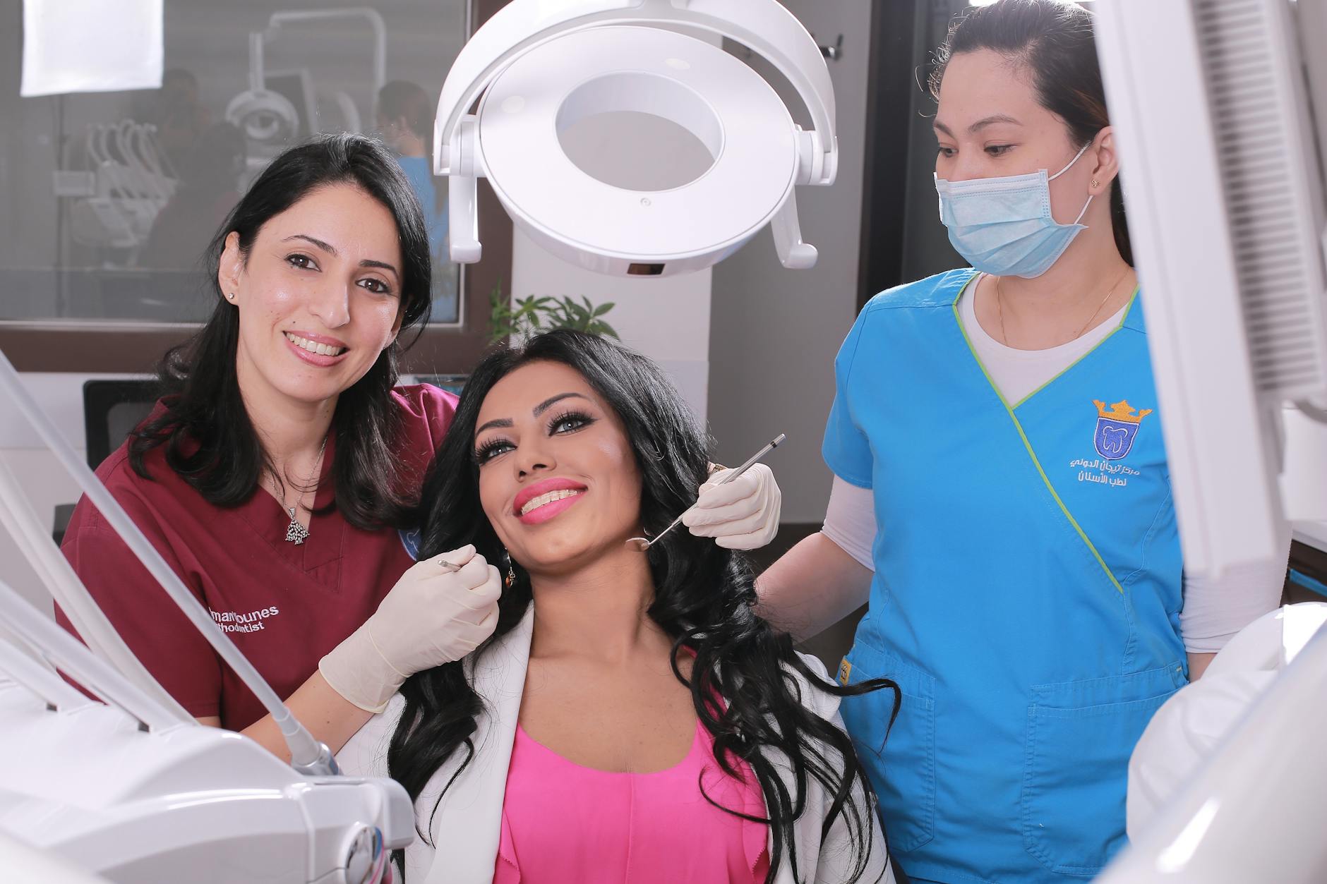
Most patients find digital face scans simple and comfortable. You look at a point while a camera captures your facial features and natural head posture—there’s no contact and no messy trays. We then match that 3D face to a scan of your teeth so the plan reflects your real smile and bite. The visit feels more like taking photos than having a procedure.
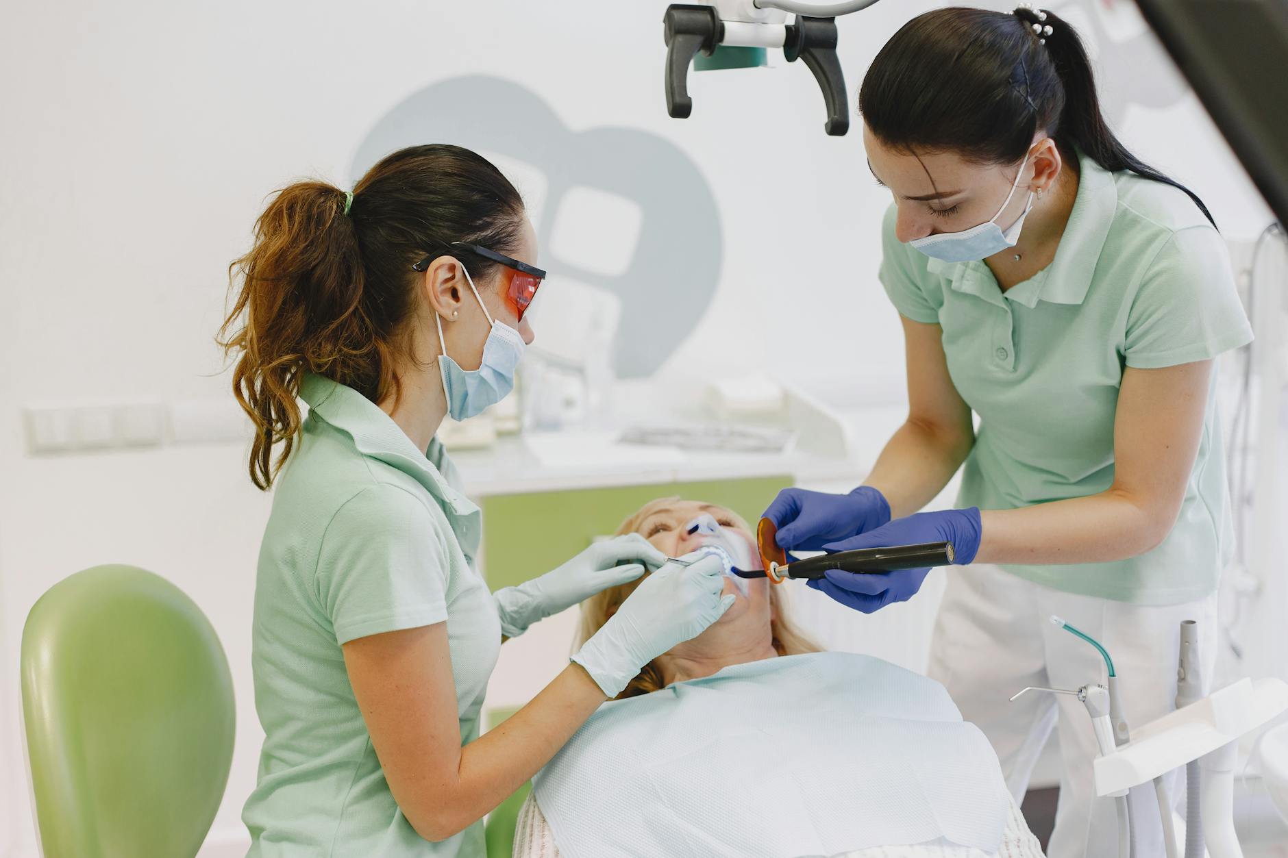
What you’ll notice: brief instructions to relax your face, a few seconds of image capture, and a small intraoral scanner that gently glides over your teeth. The face scan is non-contact; the mouth scan uses a smooth wand and pauses are easy if you need a break. Many patients prefer digital impressions over conventional putty impressions, reporting better comfort and satisfaction in comparative studies for implant restorations [7].
During the appointment, we merge your face and tooth scans on screen. This lets us align the bite plane to your eyes, center the midline, and preview tooth display at rest and in a smile. You can see options in real time and give feedback before anything is made. In the facebow vs digital face scan esthetics conversation, the digital path adds a clear, shared picture of your own features, which helps everyone agree on what looks natural for you.
If you’re prone to gagging or feel anxious, tell us. We can adjust head position, take breaks, and use small tips for the scanner; some patients also do well with gentle oral sedation options. A short bite record may be taken to sync jaw position; it sets how your teeth meet in the virtual plan. Light sounds from the scanner and camera flashes are normal. Your images are part of your dental record and used only to plan your care. Most people leave saying the process was quicker and easier than they expected, and that seeing their own face on screen made choices clearer—especially for esthetic goals like tooth length, symmetry, and smile fullness [7].
Future Trends in Digital Dentistry
Digital dentistry is moving toward connected, data‑driven planning that links your face, bite, and teeth in one virtual space. 3D face scans, intraoral scans, and a virtual articulator will work together so teams can test esthetics and function before touching a tooth. Artificial intelligence, 3D printing, and better imaging will make this process faster and more predictable for everyday care.
Virtual workflows are getting more lifelike. Expect wider use of virtual articulators that can include jaw‑motion data, helping clinicians orient teeth to your natural head position and smile with fewer physical steps. 3D face scans will likely become a standard record, improving how midlines, occlusal planes, and tooth display are set on your own image. These tools support clearer previews, easier plan changes, and safer “test drives” with printed mock‑ups before final work.
Artificial intelligence and augmented or mixed reality are expanding in dental education and are poised to support clinical planning and patient communication—think real‑time checks of symmetry, tooth length, or plane, and guided visualization for procedures [8]. In prosthetics, digital dentures and “smart” approaches—scanning, CAD/CAM design, and additive manufacturing—are redefining how complete dentures are made and refined, with better standardization from scan to try‑in [9]. Implant care continues toward data‑driven planning and guided workflows that integrate 3D face data, tooth scans, and 3D X‑rays (CBCT) to improve precision and patient‑centered decisions [10].
Multimodal records will also help unify esthetics and function. A stable natural head position, a consistent bite record, and high‑quality face and tooth scans can be captured the same way over time, making it easier to track changes and fine‑tune plans. For many cases, this reduces the need for repeated physical transfers; for complex bites, teams can still add a kinematic facebow or jaw‑motion recording for extra confidence. In the facebow vs digital face scan esthetics conversation, the future is likely a blended approach—use a clear 3D face reference for esthetics, verify function with reliable bite and movement records, then print or mill a provisional that lets you preview and adjust before finalizing.
Frequently Asked Questions
Here are quick answers to common questions people have about Facebow to Face Scan: Modern Smile Alignment in Glendale, AZ.
- What is the purpose of a facebow in dentistry?
A facebow helps dentists accurately transfer the position of your upper jaw to a mechanical jaw simulator called an articulator. This ensures that any planned dental work, such as crowns or dentures, aligns well with your bite and facial features. By referencing facial landmarks, facebows aid in setting the correct occlusal plane, midline, and tooth display, which is crucial for both functionality and aesthetics.
- How do digital face scans compare to traditional facebows?
Digital face scans capture 3D images of your face and merge them with mouth scans to visually align your smile and bite using software. Unlike facebows, they offer quick and non-contact recording and enhance detail in dental planning. Both methods strive for the same outcome: placing teeth where they look natural and function well. Modern digital scans often integrate seamlessly into workflows, allowing for easier manipulation and adjustment.
- Why are digital face scans useful for planning dentures?
Digital face scans are invaluable for denture planning because they accurately capture facial symmetry and head posture. This helps ensure that dentures fit naturally with your lips and smile. By visually integrating with the mouth scan, they guide the design process to ensure optimal lip support and fullness, which improves both comfort and appearance.
- Can digital face scans help in aligning teeth with facial features?
Yes, digital face scans can greatly assist in aligning teeth with your facial features. By merging a 3D face scan with an intraoral scan, dental professionals can visualize and adjust the occlusal plane, midline, and tooth length to fit harmoniously with your face. This improves not only the aesthetics but also the functional fit of the dental restorations.
- Are digital face scans comfortable for patients?
Most patients find digital face scans to be comfortable and non-invasive. The process involves looking at a point while a camera captures facial images, and there is typically no contact involved. Compared to traditional impressions, many patients prefer digital scans as they avoid the discomfort of messy trays.
References
- [1] Comparison of two arbitrary cast transfer systems with a kinematic facebow for mounting a maxillary cast on a semiadjustable articulator. (2022) — PubMed:33736862 / DOI: 10.1016/j.prosdent.2020.12.023
- [2] Comparison of the Kois Dento-Facial Analyzer System with an earbow for mounting a maxillary cast. (2015) — PubMed:25979448 / DOI: 10.1016/j.prosdent.2015.02.022
- [3] The Orientation in Space of the Maxillary Arch: New and Old Devices in the Prosthetic Digital Workflow. (2025) — PubMed:39473290 / DOI: 10.1111/jerd.13342
- [4] Evaluation of Smile Aesthetics in Dental Students: Perceptions of Tooth Colour Changes Due to Incisor Inclination and Micro- and Mini-Aesthetic Characteristics Assessed by Professionals and Laypersons. (2025) — PubMed:40863083 / DOI: 10.3390/dj13080380
- [5] Eco-Friendly Approaches to Enhance Dental Aesthetics and Patient Satisfactions Using Digital Smile Design: A Systematic Review. (2025) — PubMed:40895222 / DOI: 10.2147/CCIDE.S535436
- [6] Impact of material and silicone index support using additively manufactured trays on the reproducibility of diagnostic trial restorations (mock-up): A comparative in-vitro study. (2025) — PubMed:40750082 / DOI: 10.1016/j.jdent.2025.106013
- [7] Patient-Reported Outcomes of Digital Versus Conventional Impressions for Implant-Supported Fixed Dental Prostheses: A Systematic Review and Meta-Analysis. (2025) — PubMed:41003130 / DOI: 10.3390/jpm15090427
- [8] The Integration of Artificial Intelligence and Augmented Reality in Dental Education: Current Applications and Future Potential. (2025) — PubMed:40479539
- [9] From Conventional to Smart Prosthetics: Redefining Complete Denture Therapy Through Technology and Regenerative Science. (2025) — PubMed:40572792 / DOI: 10.3390/medicina61061104
- [10] The Future of Dental Implants: A Narrative Review of Trends, Technologies, and Patient Considerations. (2025) — PubMed:40970042 / DOI: 10.7759/cureus.90380