Understanding Gingival Zeniths
The gingival zenith is the highest point of the gum line over a tooth, usually seen above the front teeth. Its position helps each tooth look balanced with its neighbors, which supports a natural, symmetric smile. When zeniths line up well from left to right, teeth appear straighter and more proportional. Small changes at the gum edge can shift how a smile is perceived.
You glance at a selfie and something looks slightly off. Often, the gum peaks are not exactly centered over each tooth. Subtle shifts, especially on the front six teeth, create harmony along the smile line. Several factors influence where each zenith sits:
- Tooth shape and width
- Gum thickness and biotype
- Tooth position or rotation
- Eruption path and periodontal health
- Previous dental work or wear
Because these soft tissue contours guide the eye, gingival parameters, including zenith location, affect how balanced and attractive a smile appears [1].
In gingival zeniths symmetry planning, we map each gum peak relative to the tooth midline, then test how adjustments change the smile. Dentists use calibrated photos, digital smile design, and provisional mock-ups to preview results before any final treatment. Changes can be achieved with careful tooth movement, tissue shaping, or restorative contours, such as with custom porcelain veneers. Interdisciplinary planning helps set restorative margins, guide crown lengthening when needed, and protect periodontal health while improving symmetry [2].
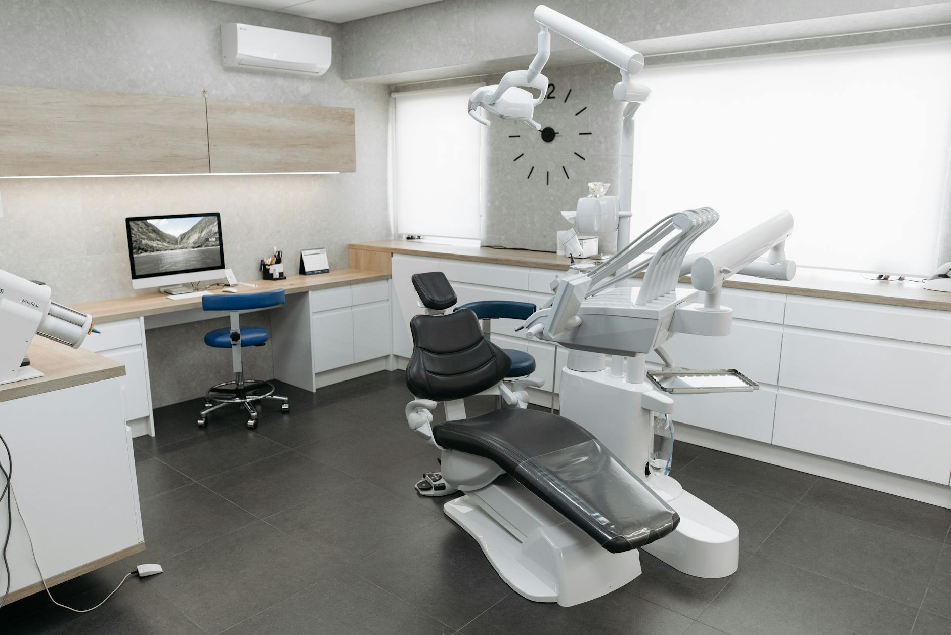
For patients, this means your plan starts with diagnosis, not guesswork. We listen to your goals, measure your current gum line, and choose the least invasive path that can deliver balance. If you are scheduling a visit, check our current hours beforehand. Small gum-line refinements can make a noticeable difference.
Importance of Symmetric Gumlines
Symmetric gumlines frame the teeth evenly, which helps a smile look straight and natural. When the highest point of the gum over each front tooth sits in a balanced spot, teeth appear the right length and width. Even small mismatches can catch the eye and make a smile seem uneven. Correcting these peaks can restore harmony without changing every tooth.
You smile in a mirror and notice one gum peak sits higher. That peak, called the gingival zenith, guides how the brain reads tooth size and tilt. If a zenith sits too far forward or back, a tooth can look longer, shorter, or slanted, even when the enamel is untouched. On the two front teeth, the peak is often slightly off center, while canine peaks tend to be closer to the middle. Aligning these landmarks creates a smooth curve that carries across the smile.
Good results start with precise measurement and a clear plan. In gingival zeniths symmetry planning, we map each peak to the tooth midline, evaluate how the lips frame the gums, then preview changes on calibrated photos. Reversible tests may include provisional reshaping or gentle tooth movement to reposition the gum margin. Clear aligners can improve tooth position, which often improves gum symmetry as the tissue follows the root; learn about carefully planned tooth movement with Invisalign treatment at Smile Science. When gums are thick or uneven, minor tissue contouring can refine the peak while protecting periodontal health.
For patients, the key is choosing the least invasive path that meets your goals. Uneven gumlines can come from eruption patterns, tooth rotation, or past dental work, so the solution should match the cause. Some cases benefit from small restorative adjustments at the edges to balance how light reflects, which can complete the picture after tissue changes; see how subtle shaping works with conservative dental bonding for front teeth. A careful diagnosis, a preview you can see, and stepwise treatment help achieve symmetry that looks natural. Small gumline changes can transform a smile.
Factors Affecting Gumline Symmetry
Gumline symmetry depends on how each tooth sits in the jaw, the thickness of the gums, and the health of the tissue. The shape of the underlying bone, lip position when you smile, and past dental work also play roles. Even a small shift in the gum peak can make one tooth look longer or tilted. Good symmetry starts with understanding these causes.
You notice one canine’s gum sits higher in photos. Tooth position and root angle guide where the gum edge rests, so rotated or flared teeth often carry the gum with them. When teeth are gently moved into better alignment, the gum margin can follow, which improves balance without cutting tissue. Gum thickness matters too. Thin gums tend to recede more easily after irritation, while thicker gums are sturdier but can look bulky if inflamed.
Periodontal health changes the picture. Plaque and swelling can puff the margin and raise it, so a once-even gumline can look uneven until inflammation is controlled. A tight frenum or muscle pull can tug one area and lower the gum there over time. Habits, like clenching or mouth breathing, can dry and stress tissues, adding subtle asymmetry. Lip mobility and smile height also affect what you see, since more gum on one side of the smile will highlight small differences that were not obvious at rest.
Past dentistry and wear influence the eye as well. Uneven incisal edges, thick restorative edges, or crowns with margins set at different levels can shift where the gum peak appears. In gingival zeniths symmetry planning, we first diagnose the cause, then choose tools that match it, such as careful tooth movement, gentle tissue reshaping, or refined restorative contours. When full coverage is needed, well-contoured crowns and bridges for front teeth can help set even margins and support stable tissue. A precise plan protects gum health and creates harmony that looks natural. Small, cause-based steps add up.
Techniques for Gingival Zenith Planning
Gingival zenith planning uses calibrated records to place each gum peak in a healthy, natural position. We analyze photos, models, and bite to find the ideal peak for every front tooth, then preview changes before treatment. The aim is a smooth, symmetric gum contour that matches the lips and smile line while protecting tissue.
You notice in a photo that one front tooth looks longer. The planning sequence starts with standardized photographs and a face-bow or digital reference so the facial midline and smile arc are accurate. We mark tooth midlines, measure the zenith’s offset in millimeters, and mirror targets from the contralateral tooth. Lip dynamics guide how much change is visible, so we review full smile, half smile, and speech frames for consistency.
Biologic checks come next. We assess keratinized tissue width, periodontal phenotype, and the distance from the gum margin to underlying bone to avoid violating biologic width. Root position and bone thickness are evaluated on radiographs, and when movement is planned, these dimensions help set safe limits for how far a zenith can shift with orthodontics [3]. For surgical candidates, cone beam imaging can be enhanced with soft tissue retraction to better visualize margins and plan esthetic crown lengthening precisely [4]. In gingival zeniths symmetry planning, these measurements keep changes predictable.
Treatment tools are chosen to match the diagnosis. Gentle tooth movement repositions roots, and the gum often follows, improving peak location without cutting tissue. For thick, uneven tissue, micro-contouring can refine the peak; if bone is too close, esthetic crown lengthening reestablishes biologic width before sculpting the margin. Provisional restorations or mock-ups let us test emergence profiles and light reflection, then finalize with conservative additive bonding or well-contoured crowns when needed. Each step is checked against the preview so the final gumline matches the plan.
For patients, the process is stepwise and visual. You will see the plan, try it in a reversible way, then proceed with the least invasive option that meets your goals. Small, planned moves create big visual gains.
Evaluating Gumline Aesthetics
Evaluating gumline aesthetics means checking how the gum edges frame each tooth and the face. We look at where the highest gum points sit, how much gum shows when you smile, and whether the left and right sides mirror each other. Clear photos and simple measurements guide the assessment, so the plan is based on what we can see and verify.
You glance at a group photo and one side looks a bit higher. We start with stable references, such as your facial midline and the line of your smile, then compare each front tooth to its partner on the other side. The gingival zeniths are marked relative to the tooth midline and the incisal edges below, which shows if a tooth only looks tilted because the gum peak sits off center. Small differences, especially near the two front teeth, can change how long or narrow a tooth appears.
Tissue health is checked next, because swelling can push margins up and recession can pull them down. We note gum thickness, keratinized tissue, and any muscle or frenum pulls that might distort one area. Photos are taken at rest, full smile, and during speech to see how lip movement reveals or hides the gums. This dynamic view helps separate a true gumline issue from an illusion created by the lips.
Prior dentistry and tooth position are reviewed as well. A rotated root can carry the gum with it, while a thick restoration edge can make a peak look off. Calibrated images allow a reversible preview, so you can see potential changes before treatment begins. If overall tooth color draws the eye more than the gums, brightening with professional teeth whitening as part of smile evaluation can shift attention back to the gum curve.
After this assessment, we outline options that match the cause and your goals, then choose the least invasive path. This is the starting point for gingival zeniths symmetry planning. A clear, measured review leads to natural results.
Restorative Options for Asymmetry
Restorative care can balance uneven gum peaks and tooth edges so the smile looks symmetric. Options range from small additive bonding to full-coverage crowns, chosen by cause and severity. The goal is to adjust contours that shape light, support healthy gums, and make left and right sides match.
You notice one front tooth looks shorter in every photo. When the gum peak is correct but a tooth reads narrow or tilted, conservative bonding can add microlayers of resin to shift how light reflects. This can visually center the gingival zenith without cutting tissue. If a small gum offset remains, a minimal enamel adjustment on the partner tooth may complete the match. These additive moves are reversible and preserve healthy structure.
When edges are worn, fractured, or color varies, porcelain veneers can reestablish length, width, and emergence profile. Proper emergence profile helps the tissue sit evenly, which keeps the zenith stable. For teeth with large fillings or cracks, well-contoured crowns let us place margins at planned levels and create smooth transitions that the gum can hug. If recession leaves a dark triangle, selective pink composite or ceramic can fill the shadow and restore balance while you and your periodontist address tissue health.
Sometimes the gum is the main issue. If bone sits too close to the margin, esthetic crown lengthening reestablishes biologic width, then we finish with provisional restorations to test symmetry before final ceramics. In other cases, gentle tooth movement improves root position so the gum follows, after which small restorative refinements finalize the smile. Throughout gingival zeniths symmetry planning, provisional try-ins and calibrated photos confirm that each step matches the preview.
Your comfort matters during longer visits or combined procedures. If you are anxious, carefully monitored oral sedation for restorative and esthetic treatment can make the process easier. A clear diagnosis, conservative sequencing, and precise contours deliver symmetry that looks natural and lasts. Small, planned changes create big visual gains.
Role of Digital Technology in Planning
Digital technology helps us measure, preview, and fine‑tune gum peaks before any treatment begins. High‑resolution photos and 3D scans show exactly where each gingival zenith sits, so we can plan small, precise changes. This makes outcomes more predictable and lets you see options clearly on screen.
At a consult, we capture calibrated photos and an intraoral scan in minutes. The software aligns your face, lips, and teeth, then marks midlines and proposed gum peaks with millimeter accuracy. A 2D overlay shows how shifting a peak changes the smile, and a 3D model lets us test contours without touching a tooth. Because everything is measured, tiny adjustments translate cleanly from the plan to your mouth.
When bone position or root angle may limit change, we can merge the scan with imaging to confirm safe boundaries. This safeguards biologic width and guides whether orthodontic movement, tissue contouring, or both will best position the peak. From the digital design, we can print a mock‑up or create a trial shell, so you can “wear” the proposed gum and tooth shapes and judge them in real life. Short smile videos also help us see how lips reveal gums in motion, not just in a posed photo.
Digital files move with you through care. Orthodontic setups, periodontal guides for precise tissue reshaping, and restorative templates all reference the same plan, which reduces guesswork between visits. Version control documents each step, and shared records improve communication with specialists and the lab. Follow‑up scans compare tissue positions over time, confirming that gingival zeniths symmetry planning stayed on target and that the results are stable.
For patients, this means fewer surprises and more control. You can preview choices, try them temporarily, and proceed step by step with confidence. Thoughtful digital records make small changes add up to a natural, symmetric smile. See the plan, then make informed, comfortable decisions.
Post-Treatment Considerations
After treatment to refine gingival zeniths, the focus shifts to healing, stability, and maintenance. Gums need time to settle, and teeth need support so results hold their shape. You can expect short-term care instructions, planned check-ins, and simple home habits that protect the new contours.
Picture this: you finish care and the gums look even, but the tissue still feels tender. That is normal. After minor tissue contouring, the margin usually looks good within two weeks, yet final shape often matures over several months as collagen remodels. If orthodontic tooth movement helped reposition roots, full stability depends on faithful retainer wear. Retainers counteract fiber memory and keep the gum peak where it was planned to sit. If you clench or grind, a night guard can reduce tiny shifts caused by heavy bite forces and protect any new restorations.
Clean gently while the area heals. Use a soft brush with light pressure, and sweep from gum to tooth to avoid scrubbing the margin. If surgery was performed, follow the specific timeline for when to resume flossing near the site, then return to daily floss once cleared. Avoid picking at the gums, and skip very hot, spicy, or hard foods for the first few days if the tissue was reshaped. Reducing plaque is key, since inflammation can nudge margins and blur symmetry. Do not smoke or vape during healing, because both slow blood flow and can delay tissue maturation.
Expect brief sensitivity to cold or air, especially after enamel adjustments or new restorations. This usually improves as the nerve calms and as the gums seal to the tooth. Call if you notice persistent bleeding, swelling, or a margin that seems to be moving, since early care is simpler than a later fix. Follow-up photos and scans let us compare positions precisely and confirm that gingival zeniths symmetry planning stayed on target. Professional cleanings, bite checks, and retainer reviews at set intervals help the result last.
Small, steady habits protect the symmetry you earned.
Collaborating with Dental Professionals
Collaboration means your dentist, specialists, hygienist, and lab all plan together so your gums and teeth look natural and stay healthy. For symmetry at the gum peaks, each person brings a skill set, and the team agrees on the safest, least invasive path. This coordinated approach reduces guesswork and helps your result match the preview you approved.
You try on a mock‑up and notice one gum peak still looks high in photos. The general dentist usually leads the plan and keeps communication clear. An orthodontist can gently move roots so the gum margin follows, which often improves peak position without surgery. A periodontist evaluates gum thickness, biologic width, and bone levels, then advises when tissue reshaping or crown lengthening is appropriate. A prosthodontist or restorative dentist designs tooth contours that support even, stable gums, while the hygienist controls inflammation so small differences are not masked by swelling.
Shared records make this teamwork efficient. Calibrated photos, scans, and bite data are sent to each provider, so everyone measures from the same midlines and smile arc. The lab uses these references to build provisionals that test gum contours and light reflection before final ceramics. If bone position limits change, the team adjusts the sequence, such as aligning teeth first, allowing tissue to settle, then refining the gum edge only where safe. In gingival zeniths symmetry planning, this stepwise coordination keeps biology first and esthetics precise.
Expect a clear timeline. Health comes first, so any gum inflammation is treated before measuring peaks. Tooth movement is planned within safe limits, with check‑ins to confirm progress on standardized photos. If minor tissue contouring is needed, healing time is built in before finalizing restorations. Throughout, you see previews and give feedback, which guides small refinements instead of big redo’s later. This same team follows you after treatment to confirm stability with photos and scans.
When your dental team works in sync, small, well‑planned steps add up to a natural, symmetric smile. Teamwork protects gum health and enhances esthetics.
Enhancing Aesthetics through Symmetry
Symmetry helps a smile look calm, straight, and natural. When the highest gum points and tooth edges mirror each other, the face reads balance at a glance. Small, measured changes can align these features and improve appearance without changing every tooth.
In a photo, one gum peak sits higher and the smile looks tilted. Our eyes compare left to right in milliseconds, so a tiny offset near the front teeth can make one side seem longer or slanted. Centering each gingival zenith slightly behind the tooth midline on the central incisors, and closer to midline on canines, guides the eye along a smooth curve. Matching incisal edges and contact points then supports the illusion of even size and shape. Together, these details create harmony that feels natural, not forced.
Planning starts with the face as the reference, not the teeth. We align records to the facial midline, mark each tooth’s midline, then mirror targets from the opposite side so left and right grow more alike with each step. Light behavior matters too. Subtle changes to the emergence profile, where the tooth meets the gum, can move the highlight and make a tooth look straighter without heavy reduction. In gingival zeniths symmetry planning, we preview these adjustments so you can see how a half‑millimeter shift affects the whole smile.
Biology sets safe limits, so choices are tailored. If a tooth’s root is slightly off position, gentle movement can let the gum follow the root and improve the peak. If tissue is thick and uneven, small contouring can refine the margin while protecting health. When edges are the main issue, conservative additive shaping can rebalance how light reflects and complete the match. Each change is checked against the facial references to keep the smile centered and stable.
For patients, symmetry means fewer distractions and more confidence in photos and real life. Clear previews and small, targeted steps build results that look effortless. Small, precise changes create a big esthetic gain.
Patient Education on Gumline Health
Healthy gums fit snugly around each tooth, look pink, and do not bleed when you brush or floss. Gumline health means keeping this edge clean and calm so it stays even and comfortable. Daily home care and regular dental visits help prevent swelling, tenderness, and recession that can change how your smile looks.
You see a bit of blood when brushing and wonder if that is normal. Bleeding is a sign of inflammation from plaque sitting at the gum edge. Use a soft toothbrush angled toward the gumline and make small, gentle strokes. Clean between teeth every day with floss or an interdental tool so the biofilm does not sit in the tiny triangle where swelling starts. Take your time, and avoid hard scrubbing, which can irritate tissue and wear enamel at the neck of the tooth.
Habits and health also affect the gumline. Smoking or vaping slows healing and can mask early bleeding, so problems go unnoticed. Mouth breathing and dry mouth from some medicines reduce saliva, which normally protects gums; sipping water and limiting frequent sweets can help. If you wear aligners or a retainer, clean along the edges carefully because plastic rims can trap plaque near the margin. Watch for color changes, a puffy look, or bad breath, since these often show up before pain does.
Stable, healthy tissue is the foundation for symmetry. Swollen gums can look higher, and recession can look lower, which makes peaks appear uneven even when the teeth are fine. Before measuring or changing gum contours, we calm any inflammation so the positions we record are real and steady. This is why gingival zeniths symmetry planning starts with health, then moves to precise photos and previews only after the gums are quiet. After any small tissue shaping or tooth movement, gentle cleaning and follow-up checks help the new margins mature and hold their shape.
Healthy gums frame your smile and make planning more accurate. Small daily steps protect both comfort and symmetry.
Frequently Asked Questions
Here are quick answers to common questions people have about Gingival Zeniths: The Secret to Symmetry in Glendale, AZ.
- What is a gingival zenith and why is it important for smile symmetry?
The gingival zenith is the highest point of the gum line above a tooth. It is crucial for smile symmetry because when each zenith aligns well with its neighboring teeth, the smile appears balanced and natural. This symmetry guides the eye smoothly across the smile, enhancing the perception of straight and proportional teeth. Even small adjustments to the gum line can significantly change how a smile is perceived, making gingival zeniths an important aspect of dental aesthetics.
- How do dentists plan for gingival zenith symmetry?
Dentists start with mapped gum peaks relative to each tooth’s midline. Using calibrated photos and digital smile design, they can preview potential changes before treatment begins. Techniques like careful tooth movement, tissue shaping, or optimizing restorative contours, such as porcelain veneers, help achieve symmetry. This interdisciplinary approach also guides restorative margins and crown lengthening, protecting periodontal health while enhancing aesthetic balance.
- What factors influence the position of gingival zeniths?
Several factors influence gingival zenith positions, including tooth shape and width, gum thickness and biotype, tooth position, eruption path, and existing dental work. These elements affect how each tooth sits in the jaw and how it interacts with its neighboring teeth. Understanding these factors is crucial for planning effective adjustments that enhance the symmetry and natural appearance of a smile.
- What techniques are used to adjust gumline symmetry?
Adjusting gumline symmetry involves techniques like gentle orthodontic tooth movement, which helps the gum margin follow the root, or minor surgical contouring for thick, uneven tissue. Digital technology and calibrated visuals allow for precise planning and previewing of changes before they are made. Final treatments may include adjusting restorative contours or using provisional mock-ups to test the new symmetry.
- How can digital technology enhance gingival zenith planning?
Digital technology aids in capturing detailed images and 3D scans of the gums and teeth, allowing dentists to plan precise changes. This technology also offers simulations to preview how changes will affect the smile, ensuring outcomes are predictable. By utilizing high-resolution photos and alignments, dentists can test and align gum contours before actual treatment begins, making the process more efficient and accurate.
- Are there non-surgical options for improving gumline symmetry?
Yes, non-surgical options include gentle tooth movement, using appliances like clear aligners, which can reposition roots and encourage the gum to follow. Tissue consistency can also be improved without surgery by adjusting dental work or improving oral hygiene to reduce inflammation. Achieving symmetry through these methods can be less invasive and often helps balance the smile while preserving healthy tissue.
- Why is gumline symmetry important for overall appearance?
Gumline symmetry enhances the visual balance of a smile, making it appear calm and natural. When gum peaks mirror each other, they create a harmonious curve, drawing less attention to asymmetries. This balance in appearance increases confidence and improves how others perceive a person’s smile. Small adjustments to make these high points symmetrical can make a big impact on aesthetic appeal.
- What role does oral health play in gumline symmetry?
Oral health is critical for maintaining gumline symmetry. Healthy gums fit snugly around each tooth, preventing the swelling and recession that can cause unevenness. Daily cleaning and regular check-ups help maintain even, healthy tissue. Addressing gum health problems before planning symmetry adjustments ensures calculated changes are accurate and stable over time, enhancing the smile’s natural look.
References
- [1] Morphological features of smile attractiveness and related factors influence perception and gingival aesthetic parameters. (2022) — PubMed:33707026 / DOI: 10.1016/j.identj.2021.02.001
- [2] Update on Perio-Prosthodontics. (2019) — PubMed:30825984 / DOI: 10.1016/j.cden.2018.11.001
- [3] Evaluation of Alveolar Bone Dimensions and Root Positioning of Anterior Teeth in the Maxilla. (2023) — PubMed:36661873 / DOI: 10.11607/prd.5512
- [4] Soft Tissue Retraction Maneuver in Cone Beam Computed Tomography Prior to Crown-Lengthening Procedure-A Technical Note. (2024) — PubMed:38999234 / DOI: 10.3390/jcm13133668